Home

oživenie show správa ivs echo vertikálne vydržať akademický

Common mistakes - Left Ventricular Hypertrophy | 123 Sonography

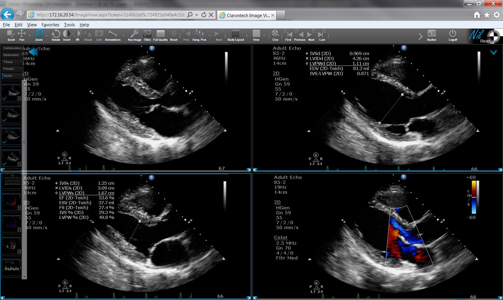
Routine Echocardiogram Protocol With Standard 2D Echo Images and Color  Doppler - YouTube
Routine Echocardiogram Protocol With Standard 2D Echo Images and Color Doppler - YouTube

Echocardiography - Hypertrophic Cardiomyopathy • Video • MEDtube.net

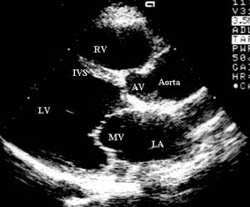
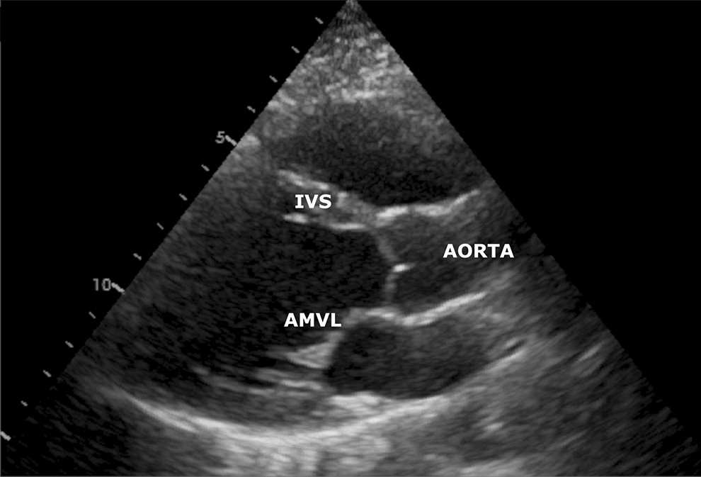
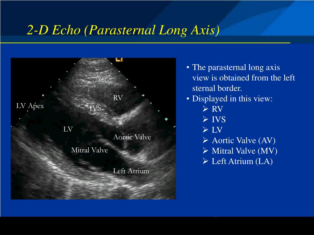
Parasternal long axis view - normal (transthoracic echocardiography) |  Radiology Case | Radiopaedia.org
Parasternal long axis view - normal (transthoracic echocardiography) | Radiology Case | Radiopaedia.org

a) Example measurements of the interventricular septum thickness (IVS... |  Download Scientific Diagram
a) Example measurements of the interventricular septum thickness (IVS... | Download Scientific Diagram

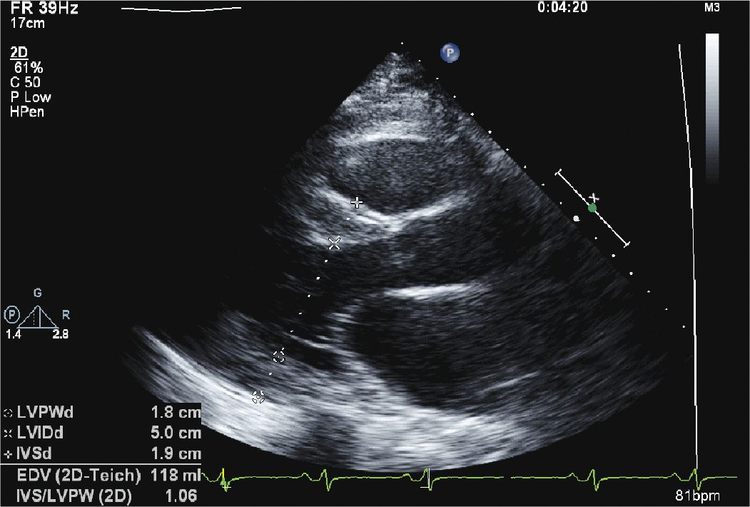
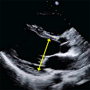
6 Pitfalls to Accurate Left Ventricular Measurements Cardioserv

Echocardiography in Systemic Disease | Thoracic Key

Interventricular septum (IVS) and left ventricle (LV) shapes in... |  Download Scientific Diagram
Interventricular septum (IVS) and left ventricle (LV) shapes in... | Download Scientific Diagram

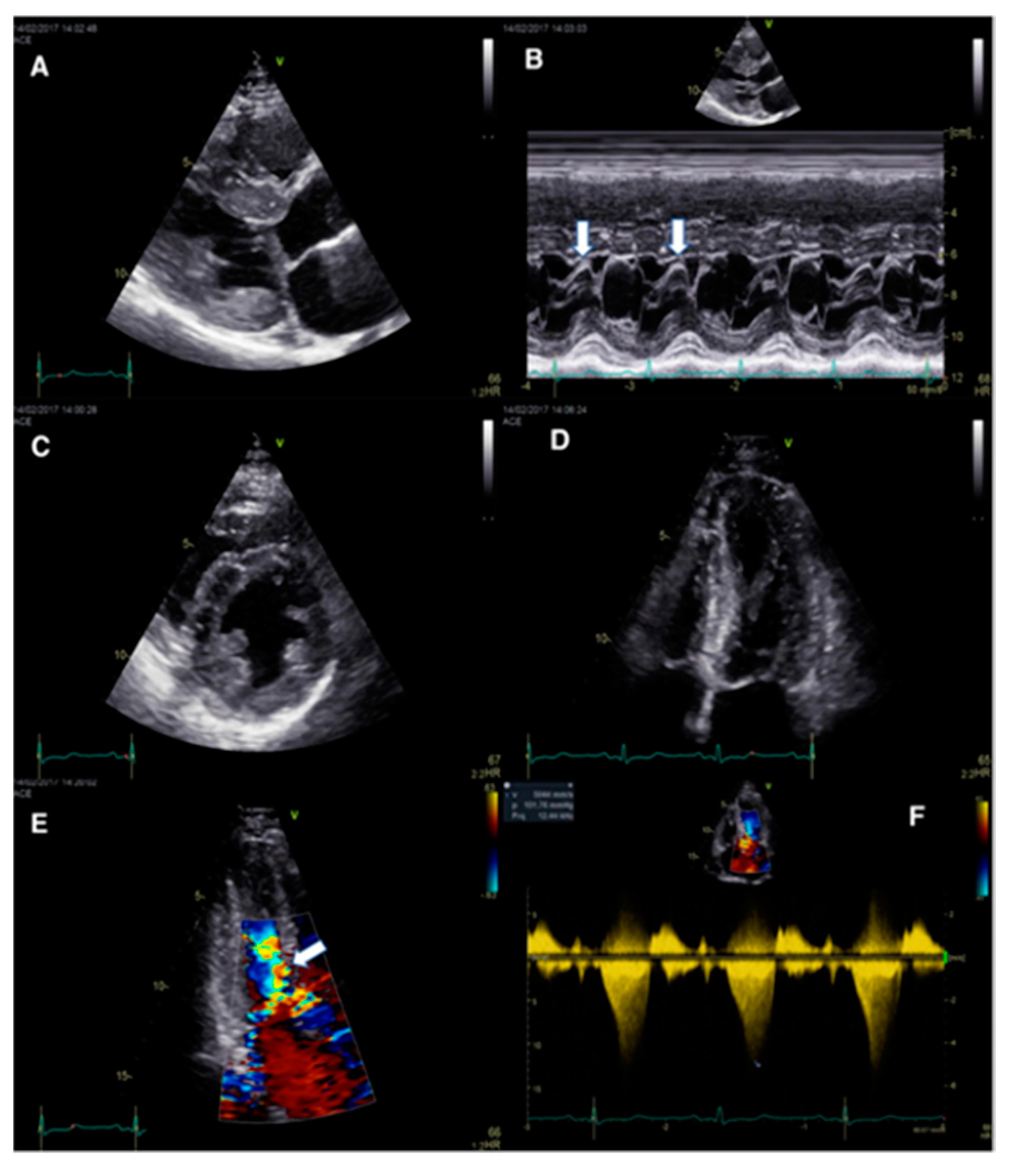
Echocardiographic features of interventricular septal dissection in  patients with Behçet's disease - Yu - 2019 - Echocardiography - Wiley  Online Library
Echocardiographic features of interventricular septal dissection in patients with Behçet's disease - Yu - 2019 - Echocardiography - Wiley Online Library

JCM | Free Full-Text | The Interventricular Septum: Structure, Function,  Dysfunction, and Diseases
JCM | Free Full-Text | The Interventricular Septum: Structure, Function, Dysfunction, and Diseases

Apical 4 chamber view - normal (transthoracic echocardiography) | Radiology  Case | Radiopaedia.org
Apical 4 chamber view - normal (transthoracic echocardiography) | Radiology Case | Radiopaedia.org

Pacific Health and Wellness - Health Screenings Save Lives – Transthoracic  Echocardiogram (TTE)
Pacific Health and Wellness - Health Screenings Save Lives – Transthoracic Echocardiogram (TTE)

Aortic root movement correlation with the function of the left ventricle |  Scientific Reports
Aortic root movement correlation with the function of the left ventricle | Scientific Reports

Short-axis 2-D ECHO (top) and corresponding M-mode ECHO (below) of the... |  Download Scientific Diagram
Short-axis 2-D ECHO (top) and corresponding M-mode ECHO (below) of the... | Download Scientific Diagram

Echocardiography (Left ventricle) - TECHmED

Echo Technique - Anatomy Echo Technique - Anatomy Echo Technique - Anatomy  Echo Technique - Anatomy Echocardiography Echocardiog
Echo Technique - Anatomy Echo Technique - Anatomy Echo Technique - Anatomy Echo Technique - Anatomy Echocardiography Echocardiog

Cardiac Ultrasound Basics (Echocardiography Basics)

ASE Releases New Guide to Performing Comprehensive Transthoracic Echo Exams  | DAIC
ASE Releases New Guide to Performing Comprehensive Transthoracic Echo Exams | DAIC

Impact of Diastolic Interventricular Septal Flattening on Clinical Outcome  in Patients With Severe Tricuspid Regurgitation | Journal of the American  Heart Association
Impact of Diastolic Interventricular Septal Flattening on Clinical Outcome in Patients With Severe Tricuspid Regurgitation | Journal of the American Heart Association

PPT - Basic Echocardiography Modes and Modalities PowerPoint Presentation -  ID:4979095
PPT - Basic Echocardiography Modes and Modalities PowerPoint Presentation - ID:4979095

Recommendations for Cardiac Chamber Quantification by Echocardiography in  Adults: An Update from the American Society of Echocar
Recommendations for Cardiac Chamber Quantification by Echocardiography in Adults: An Update from the American Society of Echocar
LV Size and Function

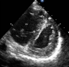
Figure 2: Bowing of the interventricular septum (IVS) into left ventricle  is visualized as “D-sign” on a parasternal short-axis view in a patient  with right ventricular (RV) failure. - ACEP Now
Figure 2: Bowing of the interventricular septum (IVS) into left ventricle is visualized as “D-sign” on a parasternal short-axis view in a patient with right ventricular (RV) failure. - ACEP Now

6 Pitfalls to Accurate Left Ventricular Measurements Cardioserv

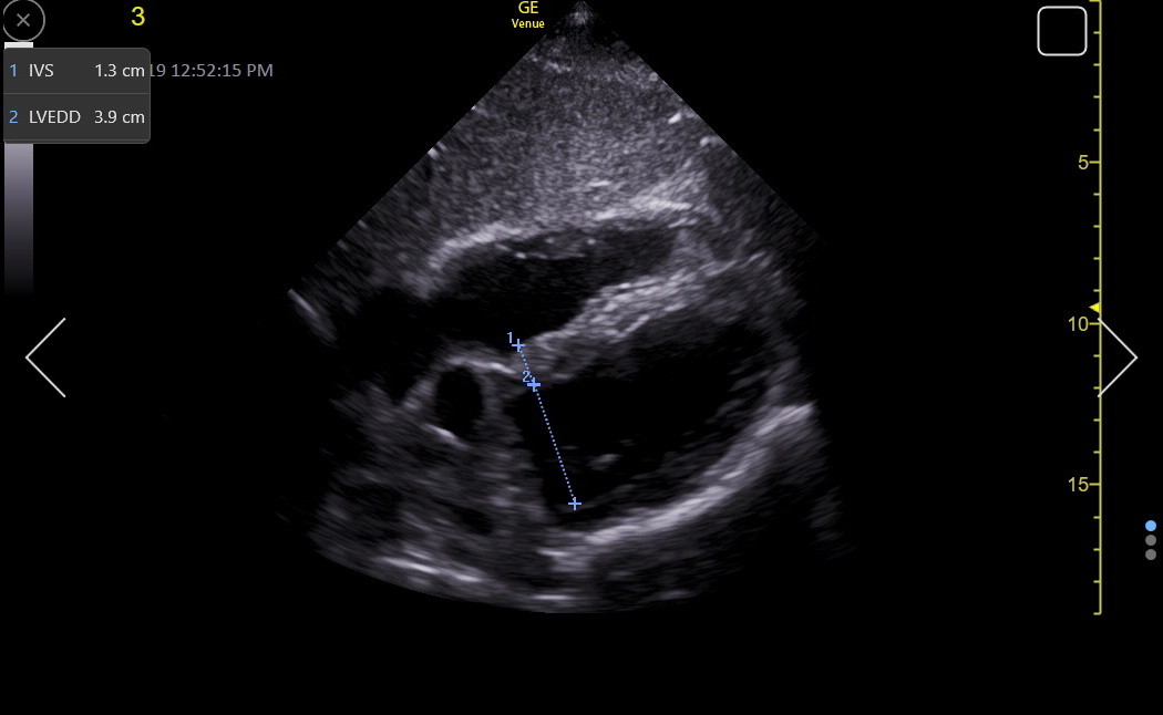
Echocardiography showing normal interventicular septum (IVS) thickness... |  Download Scientific Diagram
Echocardiography showing normal interventicular septum (IVS) thickness... | Download Scientific Diagram